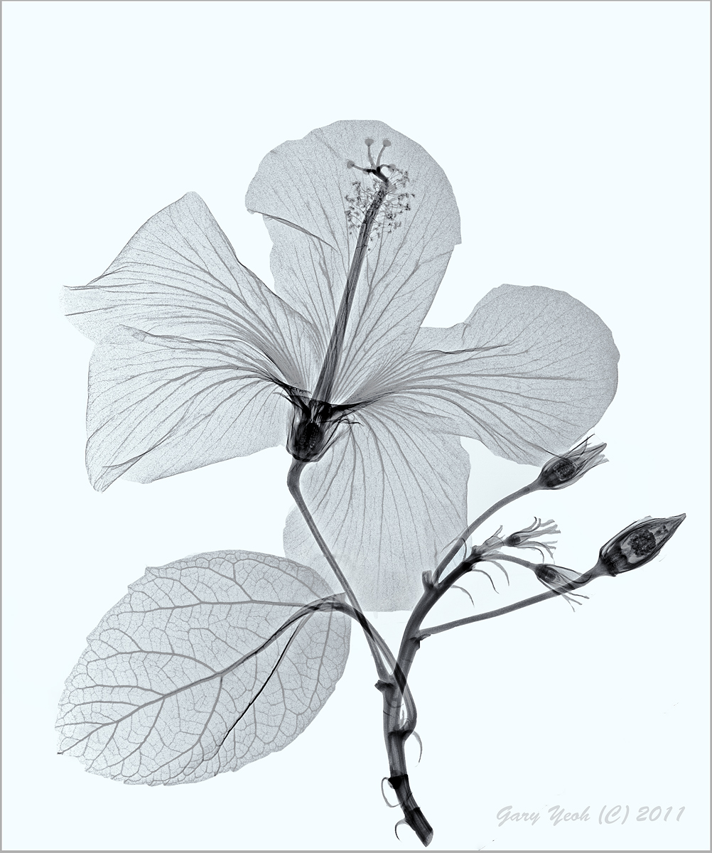
Have been working on this image for many hours to get to this stage. Boy, floral radiography is really time intensive. Hopefully it will get better as I do more of this. The first hurdle is to get a fresh flower... so I went begging. Went to the public garden near work and asked the person in charge if I could have a fresh flower to x-ray. He gave me a funny look but agreed to give me one bloom. Took it to the hospital but the x-ray department was busy so had to wait and in the mean time the flower is wilting. Finally tried to expose this with the regular plain x-ray machine at the lowest setting of 28 kV but the bg was very high. Repeat scan with the GE mammo machine set at 25 kV. The mammo machine was a new machine and the details are very good but still the petal is so thin that it is a challenge to get the selection tools to work - so had to do the hard way and manually clean up the bg.
After working on this for several hours (over many sessions), I am still mesmerised by the details of the buds and the ovules. Please see large version for details.
Dicom image processed in CS5, Silver Pro with high key & selenium toning. Posting the neg image following this and a
colorized version in Photoart.
Thanks for looking and C&C on floral radiography appreciated. The 'free trial' is about to expire and if I am thinking of doing more of these I will have to make formal arrangement to pay for the radiography.

Comments
17 commentsCompositionally, I think this would be stronger if there were no overlap in the image elements, except for the pistil and bloom. I also think more of a macro would make a very interesting image, although I have no idea though what resolution the machines are capable of. I like your processing and conversion very much. The structural detail is absolutely fascinating. I'm looking forward to the next.
pat
Hardly worth mentioning, but I'd probably crop a little off the top.
Thanks for the explanation.
D&L
This inspired me sooooo much that I grabbed an armful of flowers from the garden and drove to the local General Hospital. I showed a copy of your image to the head X-ray technician but he still called security and had me removed, now what?
Albert Darmo
Pat:
I also like the negative more but most people are more used to looking at positives and can relate to them more and that is why I have put the positive one first. The resolution of this is 1:1 - like a scan or contact print. I suppose one can do macro on this and the mammo machine can have up to 2x magnification (I think digital enlargement but in the film days the film cassette was placed further back). I am still finding my ways with this technique and I take your point about overlapping. However, unlike scan, you cannot weigh down the specimen as the washers would show up in the image - if you look very carefully at the edge of the lower petal, you can see a denser area when I have use adhesive tape to hold the stem. The strong point of radiography is the fine details that are not visible to the naked eye; and since color is not part of the equation, the shape and arrangement are the only two variables one can play with.
Scott & Bob:
I have no idea how much a x-ray machine is but it would be easily more than 100x a DSLR! However, I did a quick e-bay search for used faxitron machine which are available for US$1000-4000. This is in fact the best machine for floral photography and does not require radiation shielding of the room as it is an enclosed cabinet. I have used a faxitron machine in the 80's during my residency training at the hospital for x-raying surgical specimens (not flowers). The unit is easy to operate and does not require any specialized training - as easy as operating a microware oven! This unit can be adjusted for low kV (10-50 kV) and longer exposure of several minutes; ideal exposure level for floral radiography. I have not access to a faxitron now. The new medical machine can only go down to 25kV which is what I have to work with. The drawback of the high radiation is higher bg which is why I had to spent a lot of time manually cleaning up the bg.
Bob:
Thanks for the pointer to garden design and the fern images are certainly very beautiful. It gives me more subjects and ideas to play with.
Bill:
The x-ray department in our hospital already know I have lost my marbles.. that is why they are so accommodating
It is good to hear that you are also experimenting with floral radiography. This paper in Radiographics is quite helpful background reading. Most newer medical machines are too powerful for floral work but would work if you set the kV down as low as possible. With my calla shot, we use the setting for 'finger' which is around 40kV; the bg was a little high but the image is workable. We find the mammo a little better and the bg is cleaner. The mammo machine can go down to 22 kV. This hibiscus is shot at 25kV 40mAmps. I look forward to seeing some of your floral radiographs posts in this gallery in the near future.
I like both of them and the color version in PA gallery as well. But, I wonder how the light one would look with a dark BG?
And do you get free trial for radiography???
Great job, now we can see the normally hidden details.