Nature Photographers Network
This is the preserved archive of the original Nature Photographers Network. The community continues at naturephotographers.network.
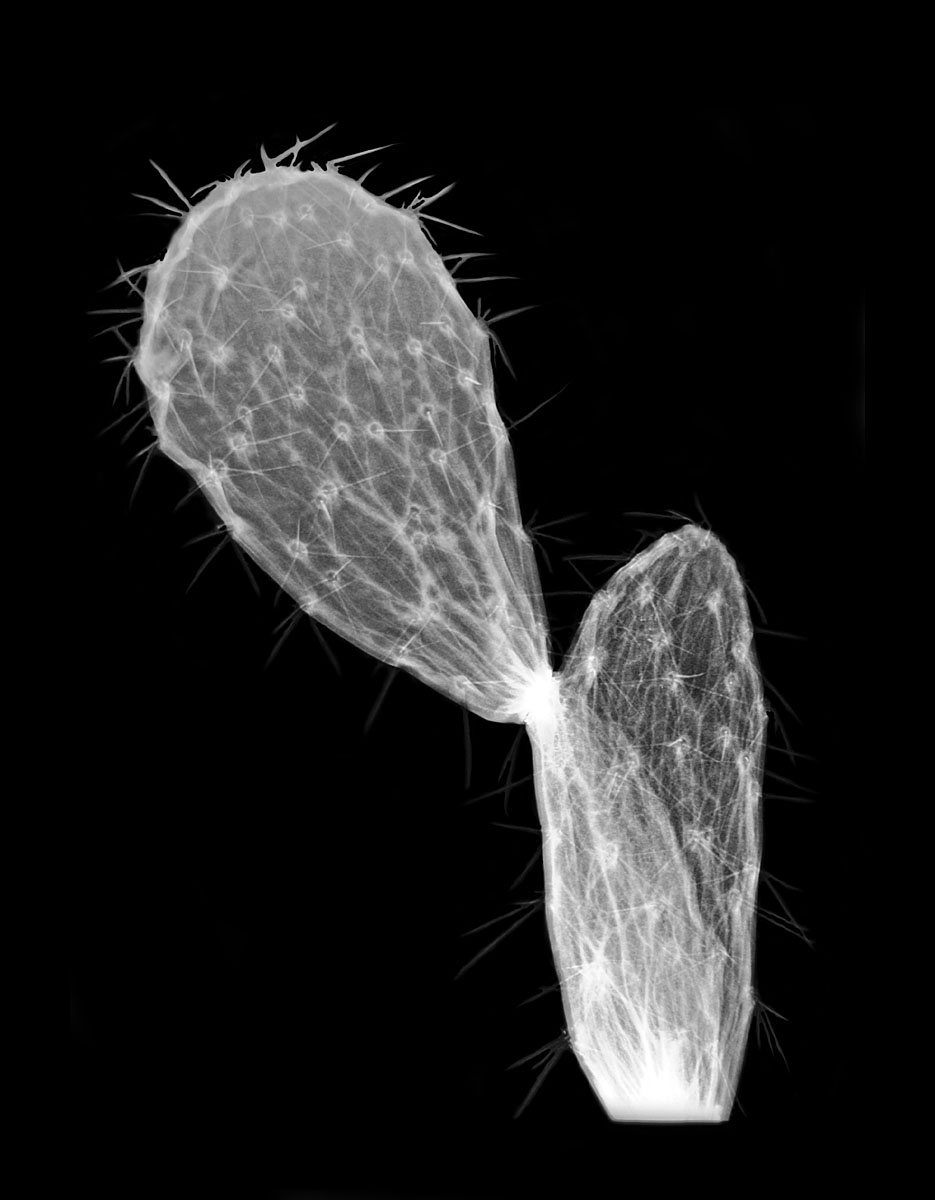
Prickly Pear X-ray
‹ PreviousPhoto Art GalleryNext ›

Prickly Pear X-ray

by Tonia Graves

Gallery: Photo ArtPosted November 2, 2011295 views9 comments
58 kVp & 2.5 mAs
Slightly dried out Desert Prickly Pear pad
X-rayed, film photographed w/ Nikon D5100 & Micro 200 on light table
Backround filled in black w/ PSE 9

For a while there, my former boss was allowing me to expirament with x-ray in my down time. She has since moved on and I fear that my new boss is not so receptive to this idea. It was fun while it lasted!

Moderators: if this doesn't fit into this category, please feel free to remove it.

Comments

9 comments
Comment posted by Rob Graham on November 3, 2011 at 10:40 PM
Nice job, Tonia. It looks like the background fill may have painted over parts of a few of the spines. FYI, this kind of image can be posted in Flora.

I'm lucky to be in the opposite position as you: my current supervisor doesn't mind me using the equipment for these images, my last manager who just left in May probably wouldn't have been as open to the idea. Do you think if you show these older images to your new boss, he/she would change his/her mind?
Registered on August 22, 2006, 842 Posts, 2824 Comments
Comment posted by Tonia Graves on November 4, 2011 at 12:38 PM
Rob, I'm not sure if I can change their minds or not. I've even offered to buy my own film! I'll keep working on it though, I think various forms of diagnostic imaging on non-human subjects yeilds some really interesting results! Btw, if you check out my portfolio I have posted a lobster x-ray as well (yes, the lobster was already dead).
Registered on October 29, 2011, 92 Posts, 1035 Comments
Comment posted by David W. Kelly on November 4, 2011 at 2:04 PM
Very nice Tonia. I hope you get to keep experimenting.
Registered on October 7, 2005, 883 Posts, 6590 Comments
Comment posted by Rob Graham on November 5, 2011 at 9:12 PM
Cool lobster in your portfolio. I should go look for some crawdad shells in the local wetlands. So you are still using film? That changes things, though if you are offering to pay for the film, then I don't see the problem. We went digital a few years ago, and finally got digital mammo and a new CT last year, so I can now "play" a bit.
Registered on August 22, 2006, 842 Posts, 2824 Comments
Comment posted by Tonia Graves on November 5, 2011 at 10:25 PM
Rob, no we are using CR but we do have film printers at some of our locations. Just out of curiosity, how do you copy the digital images from your PACS system (I realize they are all different).
Registered on October 29, 2011, 92 Posts, 1035 Comments
Comment posted by Rob Graham on November 6, 2011 at 2:15 PM
For the CT pumpkins, I burned a disc off of the workstation; same with the mammo unit I just had the mammo gal burn straight from the computer. For the few CR images I have done, I used PACS to burn a disc with the images the same way I would if I were sending a disc with a patient. I export in dicom format, though the PACS can be set to export jpgs. I am currently using the free program ImageJ to view the dicom files at home; it is a bit rough but works for what I need atm. I am glad to hear that you aren't stuck in the film ages :).
Registered on August 22, 2006, 842 Posts, 2824 Comments
Comment posted by Sam Rua on November 6, 2011 at 10:46 PM
Very cool, Tonia.
Registered on September 9, 2006, 130 Posts, 2281 Comments
Comment posted by Gary Yeoh on November 8, 2011 at 3:20 AM
Tonia:
So glad to see you posting floral radiography work! I am feeling a little lonely for a time. So interesting to see the inner vein channels in the cactus. The exposure looks great. As pointed out by Rob, some of the needles are partly covered over by the fill. It is tough to process subjects with hairs and spikes poking out of the edges. I think you could tweak the shadow and highlights a little more to get the vein to stand out more. The base and the joint look very opaque so a blend of two different kV exposures may overcome this.

Look forward to seeing more of these.
Registered on September 1, 2009, 588 Posts, 8184 Comments
Comment posted by Tonia Graves on November 8, 2011 at 1:06 PM
Gary, thank you for your suggestions. It was very difficult to get all of the spines in there, I ended up using a wacom table which helped but I may have been loosing my patience at that point! (Love to capture images, not process them). I like your idea for using 2 different exposures to maximize Kv penetration and detail. I only hope I can convince my bosses to let me do more of these from time to time!
Registered on October 29, 2011, 92 Posts, 1035 Comments