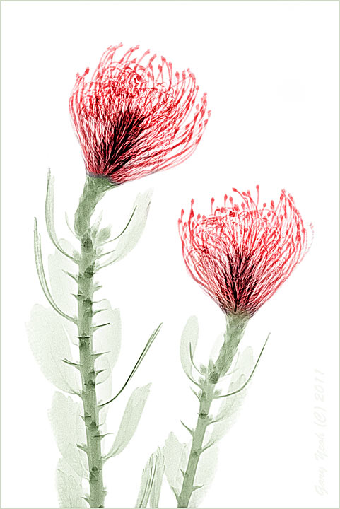
Extending image making beyond the visible light spectrum. Another x-ray image on a tango pincushion (leucospermum). We (the radiographer and myself) are still learning about flower radiography. We started this on a mammogram machine thinking that it would give better soft tissue resolution but the x-ray was not powerful enough to penetrate the dense central core. Ended up using a plain 40kV x-ray machine which was able to show the intricate pattern in the center. However, the thin leaves are all overexposed and only slightly denser than the bg. Took me a while to clean up the bg manually as the selection tools available were not good enough to define the leaves. Invert the image to black on white, adjust contrast, high pass filter, colorise in CS5. Also converted this to
color negative which I have posted in photoart.
Thanks for stopping by. Radiography is a new venture for me and the radiographer so would appreciate your comments.

Comments
15 commentsThis is very interesting. I don't really know which image I prefer most. Beautiful comp and I love the delicacy in the image elements.
I have no idea how you colorized this but I think I prefer the colorized version. I do wish there was a bit more dof.
These are both fascinating to me. I'm going to have to ponder them for a while. I'm really glad you posted these.
pat
X-ray is a different to visible light photography in that it captures incident 'light' rather than reflected light as in normal photography. There is no lens involved so there is no control of DOF (like a scanner, the object closer to the film is sharper). The DOF is much shallower than scan and in a way its a bit like pressed flowers without the compression artifact but with added 'transparent' dimension. Many people have done floral radiography and some are very nice. Dr Dain Tasker was the pioneer and probably best known. Other include Albert Richards ,Albert Koetsier and Steven Meyers.
Nathan
Albert Darmo
If I understand this correctly, when I look at the black and white image, the dark areas of the image I'm seeing are areas where the X-Rays don't pass through the subject and the white areas are transparent to the rays. The structure in the image is formed by blocking the light?
Color is not part of the process. You have no control over the dof (I'm used to that). You can't tune the subject's interaction with the light source by positioning the subject because you can't see the subject. (What you can see as solid may be opaque or even transparent to your light source.) You can't pre-visualize your image unless you've worked with the subject previously - again because the process has very little to do with what you actually see. Oh yeah, this sounds like fun. (smile)
Like Bob, I'll be very interested in seeing how this develops for you.
pat
The black on white is actually the positive of the original image. The image is formed by the object blocking the light. You pretty much sums up the experience of floral radiography. Yes, we are working in the dark at this stage and feeling our way around. If you read Steven Meyers statement, he had done about 3000+ and most of his 'failures' are due to composition. Subject selection and arrangements are paramount as there is not much control in the exposing part. Hopefully I can apply a bit of what I have picked up in Ikebana here.
Bob:
I guess we could do multiple image exposures and blend them in PS like Harold Davies high key images to get the stem transparent but so far we have only done 2 images and we have a long learning curve ahead with this technique. Another variation is perhaps multiple exposure on the same x-ray plate but varying the angle of the ray slightly (a bit like tomography) but I don't know if this is feasible. Something for the future when we get bored.
Nathan:
The radiology dept is very supportive and the staff are quite keen to learn what else they can do with x-ray.
although i dont understand the technique u've used i love the result u've got
JohnR