A Kaleidoscope of Colors Through a Microscope...
Text and photography copyright Thomas L. Webster. All rights reserved.
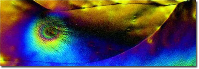
Photo 1 - Vitamin C photographed with crossed-polarized lighting displays stunning kaleidoscopic colors.
|
For the past few months I have taken my love of macrophotography into the realm of the truly microscopic, making images through a 1960s era Nikon microscope and posting those images in the NPN Macro and Close-up Gallery. As responsive as our members have been towards my previous photomicrographs, very few of my image series have been as popular as the series of vitamin C crystals photographed in crossed-polarized light. In nearly all of the responses to my posts the statement has been made, "I don't know how you do this but...". I thought this month I would demonstrate how simply these images may be made.
The Subject...
Vitamin C is an essential vitamin performing an important anitoxidant role that helps neutralize free radicals produced as a result of cellular metabolism. In the simplest of terms, free radicals are oxygen-bearing chemicals of which the oxygen has been stripped of some electrons. These free radicals can damage proteins (DNA included), lipids (fats), and nucleic acids (the building blocks of DNA and RNA). Vitamin C plays a role in directly mitigating the effects of free radicals as well as repairing vitamin E, an even more powerful antioxidant. Vitamin C is important in maintaining a healthy immune system and is absolutely essential in the formation and repair of collagen, an important ingredient of skin and connective tissues.
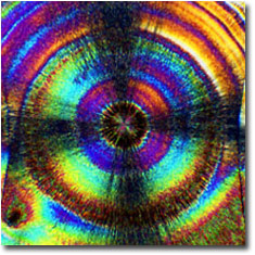
Photo 2
|

Photo 3
|
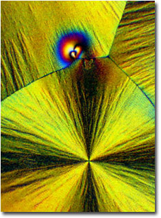
Photo 4
|
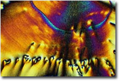
Photo 5
|
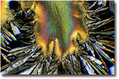
Photo 6
|
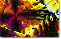
Photo 7
|
Vitamin C is water soluble and easily flushed from our bodies. Oddly enough, primates, guinea pigs, and humans are the only animals that cannot synthesize their own vitamin C. We must eat a diet rich in vitamin C to maintain our health. Citrus fruits have the highest concentrations of vitamin C but leafy greens, such as turnip greens and spinach, also have ample concentrations of vitamin C. Green peppers, strawberries, tomatoes, broccoli, turnips, sweet and white potatoes, and cantaloupe are good sources, too. British sailors in the 1600s and 1700s suffered terribly from scurvy during long ocean voyages. Through the studies of Dr. James Lind it was found that having the sailors drink lime juice prevented the onset of scurvy and, to this day, British sailors are known as "Limeys".
The Equipment and Methods...
The equipment required to view crystals under crossed-polarized light need not be elaborate. A relatively simple microscope fitted with a couple of inexpensive polarizing filters will suffice. There are many microscopes that are designed just for polarized light observations but these microscopes can run into the tens of thousands of dollars. Unless you are making accurate scientific studies of crystals, a simple set up, such as mine, will provide hours of visual and photographic enjoyment.
At first glance my microscopy setup may appear complicated. In reality, this equipment only cost a few hundred dollars and is very easy to set up and use. There is a plethora of serviceable microscopes from the 1960s and 1970s on the used equipment market. Any of these may be put to good use observing and photographing everything from crystals to "micro-critters" to histological specimens. The microscope, pictured here, is my beloved Nikon S-K trinocular microscope which was built some time in the early 1960s.
Crossed-polarized lighting requires the vitamin C crystals to be situated between two polarizing filters (see Photo 3). I place a 49mm linear polarizing filter (A) on top of the centering condenser below the slide stage. I can rotate this polarizing filter in relationship to the second polarizing filter to obtain the the maximum amount of crossed-polarized lighting. Light transmitted through the centering condenser is singly polarized before being focused onto the vitamin C crystal by the sub-stage condenser. Light passes through the vitamin C crystal (B), is imaged by the objective lens (C), and transmitted to the trinocular viewing head (D). I can view the crystals in singly polarized light and, although I cannot see the colors as yet, areas of strong refractivity will take on an obvious three dimensional appearance. The image is also diverted into the trinocular photgraphic port (E, hidden inside the microscope adapter) and is imaged onto the film plane by a special projection ocular lens. The macro-bellows also contains the second polarizing filter (F), referred to in microscope terms as the "analyzer". A 49mm polarizing filter fits perfectly flat in a recess in the front standard of the bellows.
Exposure is controlled automatically by the Canon EOS A2E camera while in aperture priority mode. Most often I shoot with Fujicolor® color print films, both for economy and for increased color saturation. It is not unusual for me to shoot from 200 to 500 exposures in a single session. To isolate the microscope from camera shutter vibrations, the camera, macro-bellows, and microscope adapter are mounted on an old enlarger support. The microscope adapter is just slightly larger than the trinocular port and, when the trinocular port is carefully centered in the adapter, isolates the camera from the microscope. The right-angle viewfinder adapter provides a convenient viewing angle and allows for focusing the image.
The macro-bellows allows for varying image magnifications at the film plane without having to change to higher power projection ocular lenses. High powered projection oculars yield "empty magnification". The amount of detail seen in a photomicrograph is regulated by the quality of the objective lens. Optical performance is always best if the eyepiece lenses and projection lenses are of the lowest practical powers for the desired image magnification. Using the macro-bellows to increase the distance between the film plane and the projection ocular lens increases image magnification while preserving details in the image. After the light rays are focused on the back plane of the projection lens the image is then focused onto the film. Focus is achieved by moving the specimen stage closer to or farther from the objective lens via the focusing knobs.
Nothing could be easier to make than microscope slides with vitamin C crystals. The pure form of vitamin C is ascorbic acid and is readily found in health food stores. As already mentioned, vitamin C is water soluble and I dissolve about 1/8th of a teaspoon of ascorbic acid in approximately 30 cc of water to make a vitamin C solution. This solution is then thinly spread on a blank microscope slide and allowed to air dry. Left on its own to dry, vitamin C will form large, circular, plate-like crystals. You can create some interesting crystal formations by scratching the surface of the forming crystals with a needle, by placing the half formed crystals in the microwave oven, or by freezing the half-formed crystals for a few hours. There really seems to be no end to the types of crystal formations that can be induced.
The Physics...
Light rays are composed of waves of light that vibrate in all planes. When light rays pass through a polarizing filter only those light rays that are vibrating parallel to the privileged direction of the polarizing filter are able to pass through the filter. This results in light rays that are all vibrating in the same plane of travel. The more oblique the light rays that strike the front surface of the filter the more these oblique light rays are filtered out. Perhaps you have noticed in your own photography that polarizing filters have their greatest effect on a landscape photograph when the scene being photographed is oriented 90° to the sun. In this orientation, the polarizing filter is blocking out the majority of oblique light rays that contribute to reflections and flare and thereby increases the saturation of the photograph.
Polarizing filters come in rotating mounts that allow the photographer to orient the privileged direction of the polarizing filter to obtain the highest degree of polarization. If the photographer were to "stack" two polarizing filters together, view a strong light source through the stacked filters, and then rotate the filters, the photographer would see that the light source would become dimmer and dimmer as the privileged direction of the two polarizing filters approach 90° in relationship to each other. Theoretically, when the privileged direction of the two filters cross at 90°, all light passing through the filters will blocked (extinguished) and the photographer would no longer be able to see the light source through the filters. In this orientation, the polarizing filters are said to be crossed, hence the term crossed-polarization. In reality, however, due to the quantum nature of light, a faintly visible and blue-shifted light source can still be seen. For photographic purposes, though, the extinction of the light is complete enough.
If all of the transmitted light is being extinguished by the crossed-polarizing filters, then how can a photographer photograph crystals under the microscope? As regards polarized light, crystals come in two "flavors". When struck by strongly polarized light, isotropic crystals allow transmitted light to pass through the crystal relatively unaltered. Since the second polarizing filter is crossed 90° in regards to the first polarizing filter, a photographer would not be able to view these crystals with crossed-polarizing filters. Anisotropic crystals, on the other hand, act as tiny prisms that break up transmitted white light into its constituent wavelengths of red, blue, green, yellow, and violet light. Uniquely, anisotropic crystals not only break up white light into its constituent wavelengths but anisotropic crystals rotate the constituent wavelengths 90° to plane of the polarized light transmitted through the crystals. The constituent light rays are then able to pass through the second polarizing filter and expose the film in the camera. The results are truly kaleidoscopic!
The Results...
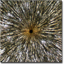
Before...Vitamin C with single polarized light (200x).
|
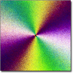
After...Vitamin C with crossed-polarized light (200x).
|
About the Images...
All of the images in this article were photographed with the microscope described above. Exposures ranging from 1/2 second to 7 seconds were recorded on either Fujicolor Super HQ ISO 100 or ISO 200 color negative film. An 82B color correction filter is added to the light path below the centering condenser to bring the tungsten illumination up in temperature to daylight illumination temperatures. The negatives were scanned on an Epson 1640 SU PHOTO flatbed scanner with a transparency adapter and final image adjustments were accomplished with Adobe Photoshop® 6.0.
- Photo 1 - 4x LOMO semi-plan achromatic objective lens with 5x Jena projection ocular + bellows extension, 50x
- Photo 2 - 20x LOMO flat-field achromatic objective lens with 5x Jena projection ocular + bellows extension, 200x
- Photo 3 - Nikon S-K microscope manufactured sometime in the early 1960s
- Photo 4 - 4x LOMO semi-plan achromatic objective lens with 5x Jena projection ocular + bellows extension, 50x
- Photo 5 - 10x LOMO flat-field achromatic objective lens with 5x Jena projection ocular + bellows extension, 100x
- Photo 6 - 20x LOMO flat-field achromatic objective lens with 5x Jena projection ocular + bellows extension, 200x
- Photo 7 - 10x LOMO flat-field achromatic objective lens with 5x Jena projection ocular + bellows extension, 100x
Author's Notes...
If you would like to see more vitamin C crystal photography visit On Closer Inspection... While you are there visit some of the other image galleries to see what is possible with modestly priced microscopes. Also, if you want to view the ultimate in vitamin images, surf on over to Molecular Expressions' Vitamin Gallery. Those images are stunning!
Tom Webster - NPN 480
Comments on NPN macro photography articles? Send them to the editor.


|